Projects & Resources
We have a number of projects on the go, and this page outlines some of the main areas of research at present. We are also passionate about developing and broadly sharing tools and technologies that we develop for our research. If these research areas and tech are of interest to you, either to come and work in our team or for collaboration, please get in touch.
Featured projects
In a 3-year project funded by New South Wales Medical Research, we are developing single molecule imaging and spatial omics tools to study systems of key signalling molecules and their regulators in the healthy and failing myocardium. We are bringing together multiplexed super-resolution imaging, big data pipelines and machine learning to understand how these molecular systems of signalling lose their balance as a heart begins to fail.
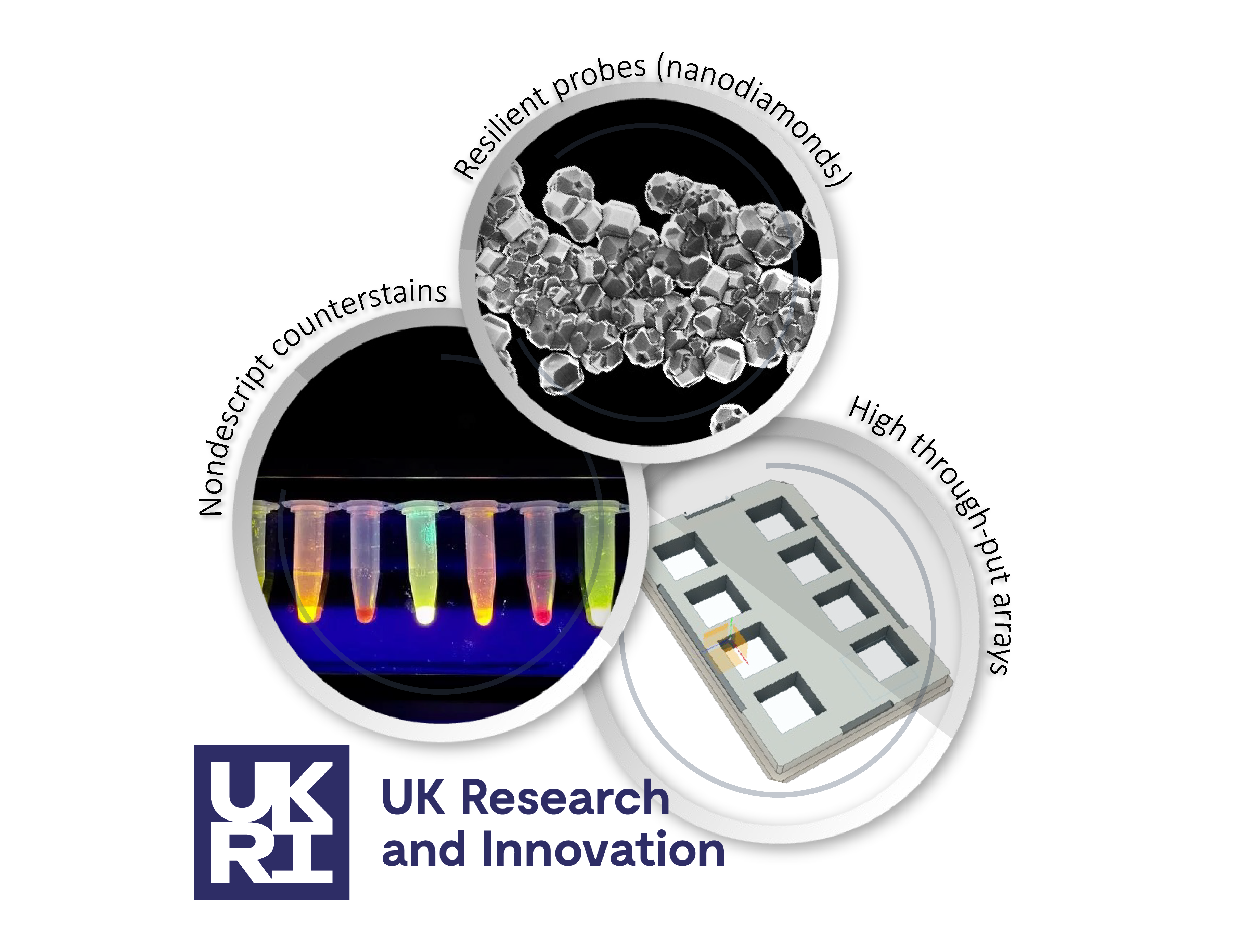
In a project originally started with a UK Research & Innovation Future Leader Fellowship awarded to Izzy, we continue to develop several innovations that are aimed at making super-resolution microscopy, expansion microscopy in particular, accessible and affordable for life sciences researchers. Amongst some of the work completed so far, we have characterised a series of 14 nondescript stains and 3D printed microplates for expansion microscopy. As a part of this project, we are also applying super-resolution to sample types and sub-fields that have not utilised super-resolution as much as mainstream cell biology. They include imaging thick tissues and whole organisms such as sea urchin larvae.

We have an ongoing interest in the Rab GTPases and their associated functions in driving the maturation and mobilisation of intracellular compartments such as endosomes and secretory granules. In a recent study published in ACS Nano, we show how we can map the nanoscale assemblies of Rab GTPases on surface sub-domains of such organelles. In ongoing work, we look to understand their relationship to calcium signals that initiate their mobilisation.